Osteoarthritis is a widespread condition in our society and stands as the most common chronic disease among people over 65 years of age, surpassing even high blood pressure and diabetes. According to the Australian Institute of Health and Welfare, around 2.1 million Australians were living with osteoarthritis in 2022. It is a significant health issue, accounting for 3% of all general practitioner visits nationwide.
Historically, there has been a gap between evidence-based guidelines for osteoarthritis management of the knee and hip and their application in clinical practice. This disconnect has often resulted in suboptimal outcomes for patients. Recognising this, an education and exercise program called GLA:D (Good Living with OsteoArthritis, Denmark) was introduced in 2014 to bridge this gap. Designed to translate evidence into practical management strategies, GLA:D aims to improve patient outcomes and quality of life.
This blog will help you understand what osteoarthritis is, its risk factors, diagnostic protocols, and various management options available to address this condition.
Knee & Hip Joint Anatomy
The knee joint is a complex hinge joint which connects the thighbone (femur) to the shinbone (tibia). It is supported by several key structures, including bones, cartilage, ligaments, tendons, and muscles, all working together to provide stability, mobility, and strength. The end of the bones is covered by the articular cartilage, a smooth, white tissue that reduces friction and absorbs shock during movement. Between the two bones sits the menisci. The medial meniscus is located on the inside of the knee, while the lateral meniscus sits on the outside. The menisci is a fibrocartilaginous cushion which distributes weight and provides further shock absorption. Encasing the joint is the articular capsule, which contains synovial fluid—a vital lubricant that reduces friction and nourishes the knee’s internal structures. Stability of the knee joint is maintained by four primary ligaments: the anterior and posterior cruciate ligaments (ACL and PCL), which control forward and backward motion, and the medial and lateral collateral ligaments (MCL and LCL), which stabilise the inner and outer knee. Tendons like the quadriceps tendon and patellar tendon connect muscles to bones, with the quadriceps muscles extending the knee and the hamstrings flexing it.
The hip joint is a ball-and-socket synovial joint that connects the thighbone (femur) to the pelvis, providing both stability and mobility essential for weight-bearing and movement. The acetabulum of the pelvis forms the socket, while the rounded femoral head serves as the ball, with both surfaces covered in smooth articular cartilage to reduce friction and absorb shock. Surrounding the socket is the acetabular labrum, a fibrocartilaginous ring that deepens the joint, enhances stability, and helps retain synovial fluid. Similarly to the knee joint, the hip joint is encased within a strong fibrous capsule lined with a synovial membrane, which provides lubrication and nutrients. Movement at the hip is controlled by various muscle groups, including the hip flexors, extensors, abductors and adductors.
Osteoarthritis – What Is It?
Osteoarthritis (OA) can affect any joint in the body where cartilage covers the ends of the bones. The joints most commonly affected are the hands and fingers, knees, and hips.
Osteoarthritis impacts the entire joint, including the articular cartilage, joint capsule, ligaments, muscles, and underlying bone. While it is often associated with older adults, it can also affect younger people, especially those who have suffered from previous injuries.
Historically, osteoarthritis was believed to be the result of simple wear and tear, leading to the misconception that joints simply “wear out” over time. As a result, the condition was often referred to as “bone on bone” or a “wear and tear disease”. However, this outdated view has contributed to the false belief that people with osteoarthritis should avoid physical activity.
In reality, research has shown that cartilage requires moderate load through physical activity, such as walking, to remain healthy. Additionally, factors such as stress, thoughts, and beliefs can influence the perception and intensity of pain in individuals with osteoarthritis, alongside the physical changes in the hip and knee joints.
Risk Factors
There are several risk factors contributing to the development of osteoarthritis, and the likelihood of developing it increases with the number of these risk factors you have. These factors can be categorised into two groups: non-modifiable factors, which we cannot change, and modifiable factors, which we can influence to reduce the symptoms of osteoarthritis.
Non-modifiable risk factors include age, genetics and sex. The risk of developing OA increases with age, particularly after the age of 50, as cartilage naturally deteriorates over time. Genetics also play a significant role; if osteoarthritis runs in your family, you are more likely to develop it. Women, especially after menopause, are at a higher risk of developing knee and hip OA compared to men, likely due to hormonal changes that contribute to this increased vulnerability.
Modifiable risk factors for developing OA in the knees or hips include joint overuse, obesity, physical inactivity, weak muscles, and poor posture. Repetitive stress on the joints, whether from high-impact activities or physically demanding work, or past injuries such as fractures of ligament damage, can accelerate joint stress and increase the risk of OA. Carrying excess weight places additional strain on weight-bearing joints, while also promoting low level systemic inflammation, which can worsen the condition. A sedentary lifestyle can lead to muscle weakness and reduced joint mobility, both of which contribute to OA. Strengthening the muscles around the joints helps stabilise them and reduce the strain on joint. These risk factors are within an individual’s control and can be managed through lifestyle changes such as maintaining a healthy weight, staying active, and avoiding joint overload.
Diagnosis
Current clinical guidelines suggest that imaging is no longer required for diagnosing knee or hip osteoarthritis, as imaging often does not correlate well with the presence or severity of symptoms. The link between changes seen on scans (such as X-rays and MRIs) and the symptoms experienced by patients is generally weak. For example, about half of people with osteoarthritis pain show no visible changes on imaging. Conversely, changes that suggest osteoarthritis, such as reduced joint space, osteophyte formation, and increased bone density beneath the cartilage, can often be seen in people who do not have symptoms. In fact, only about half of those with imaging changes actually experience symptoms.
Instead, a diagnosis should be based on a patient’s medical history, symptoms, risk factors, and findings from a clinical examination. Common symptoms of osteoarthritis include joint pain, morning stiffness lasting 30 minutes or less, and swelling. Interestingly, these symptoms can often be present for 10 to 15 years before any noticeable changes appear on imaging. Therefore, a thorough clinical examination allows for an early diagnosis, enabling earlier treatment and better management of the condition.
Management of Knee and Hip Osteoarthritis
Clinical management guidelines worldwide unanimously agree that all individuals seeking care for knee and/or hip osteoarthritis should be offered exercise therapy, education, and weight management (when necessary) as first-line treatment. Strong evidence supports these three pillars as the foundation of effective care.
Other treatments, including oral medications, manual therapies (such as soft tissue massage, joint mobilisation, and dry needling), orthotics, taping, and injections (e.g., cortisone or platelet-rich plasma), have varying levels of supporting evidence. While these interventions may provide short-term symptom relief, they offer little long-term benefit and should only be used as adjuncts to first-line treatment.
Surgery is considered the final tier of treatment and should be reserved for individuals who have exhausted first- and second-line options without achieving satisfactory outcomes. In general, knee and hip arthroscopies provide minimal benefit beyond that of a placebo or sham surgery and are therefore not recommended. In carefully selected candidates, joint replacement surgery can be highly effective, but it should be pursued only when absolutely necessary. It is important to understand the risk that comes with any surgical procedure, including infection, deep vein thrombosis, and pulmonary embolism. Additionally, since joint replacements typically require eventual revision, the goal is to delay the initial procedure as long as possible in order to minimise the number of surgeries a patient undergoes throughout their lifetime. Research also shows that patient outcomes improve with prehabilitation before surgery, so regardless of whether surgery is the chosen route, initiating an exercise program beforehand is beneficial.
The GLA:D Program is an internationally recognised education and exercise initiative designed for individuals living with symptomatic hip and knee osteoarthritis. Developed in Denmark in 2013, GLA:D was created to close the gap between recommended clinical guidelines and actual osteoarthritis care in practice.
Following its success across Denmark, the program gained international attention and was introduced in Australia in 2017 by La Trobe University’s Sport and Exercise Medicine Research Centre. GLA:D is now offered in every state and territory, including here at Bend & Mend.
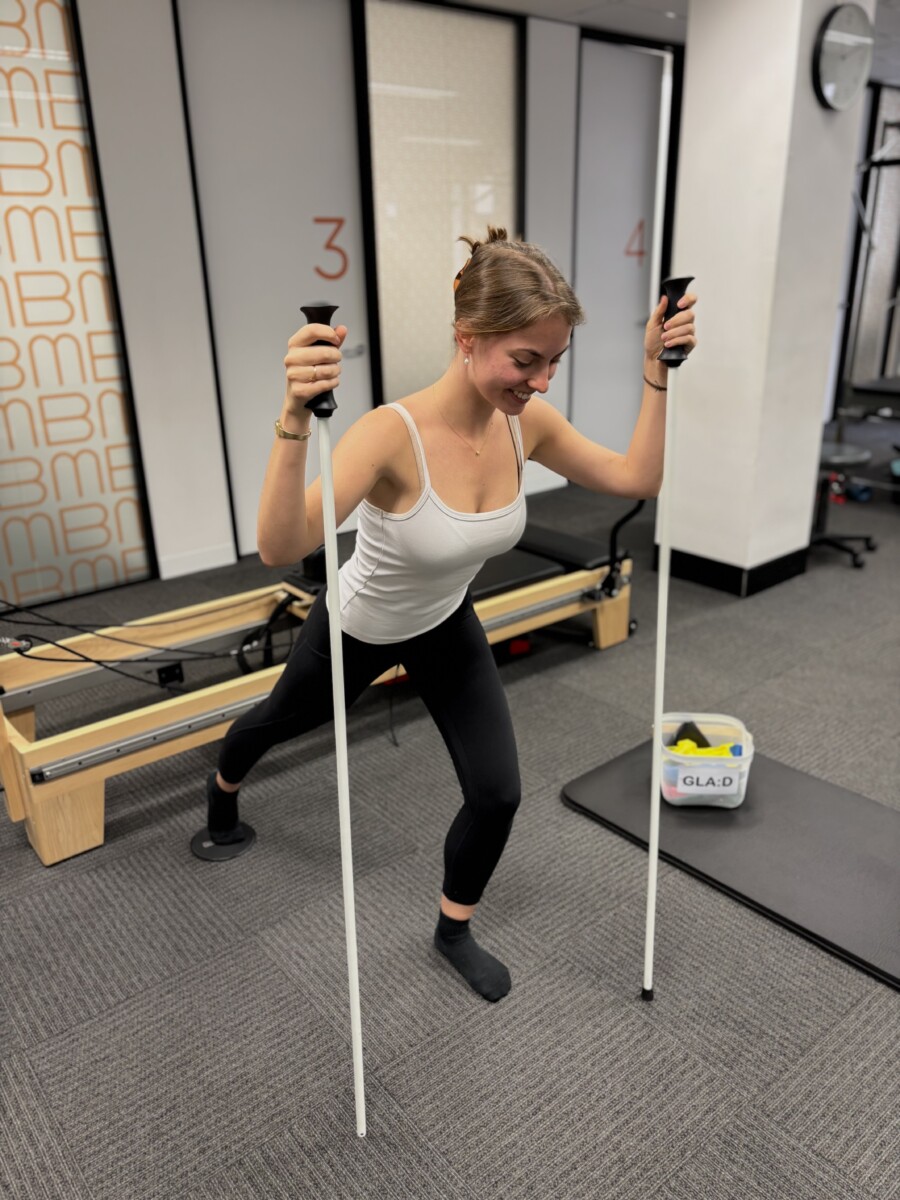 Unlike conventional osteoarthritis management, GLA:D combines both education and exercise in a structured six-week program. Participants attend two sessions per week for exercise and take part in two group information sessions. These sessions help individuals better understand osteoarthritis, including its causes, risk factors, treatment options, and the science of pain, while also providing practical strategies for self-management.
Before beginning, each participant undergoes an individual assessment with a physiotherapist to discuss symptoms, set goals, and complete baseline physical tests. A follow-up assessment is completed three months later to monitor progress and revisit initial goals.
Anyone experiencing symptoms of hip or knee osteoarthritis is welcome to participate—regardless of age, level of pain, functional limitations, or previous joint surgeries.
Those who complete the GLA:D Program commonly report reduced pain, improved mobility and physical function, decreased use of pain medication, fewer sick days, and an overall improvement in quality of life.
If you’re experiencing hip or knee pain or would like to learn more about how GLA:D can help, please get in touch with our friendly reception team at Bend + Mend.