Before we delve into what to expect in the postnatal period, it is important to first understand the fundamentals of our anatomy, particularly the pelvic floor and abdominal muscles, and the physiological changes they undergo during pregnancy. This will help you to better understand the changes that your body will undergo in the postnatal period.
Anatomy of the Pelvic Floor
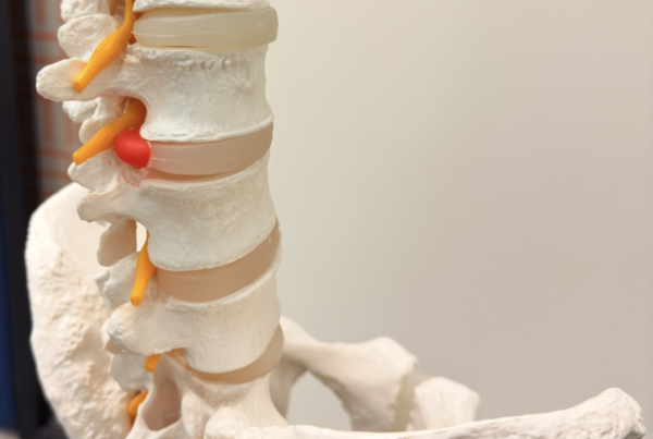
The pelvic floor, commonly referred to as the hammock of the pelvis, is a complex group of muscles, ligaments and fascia spanning from the tailbone and the pubic bone. It forms the base of the pelvis and provides crucial support for organs such as the bladder, bowel, uterus, and vagina. Working together with the deep abdominal, back, and diaphragm muscles, it stabilizes the spine and regulates intra-abdominal pressure. Additionally, the pelvic floor includes the perineum, a stretch of tissue between the vaginal opening and anus, typically measuring 2 to 5 centimetres in length.
Think of the muscles of the pelvic floor like any other voluntary muscle in our body, such as our bicep muscle. They provide ‘active’ support to our pelvic organs, allowing a woman to consciously contract and relax them. The primary muscles responsible for upward support within the pelvic floor are known as the levator muscles. Contraction of the pelvic floor muscles elevates the pelvic organs and tightens the openings of the vagina, anus, and urethra. Conversely, relaxation of these muscles allows the organs to descend slightly and facilitates the passage of urine and faeces. In contrast, ligaments and fascia provide passive support and are not within voluntary control. With time and exposure to activities that exert frequent downward pressure on the pelvic floor, such as constipation, coughing, and pregnancy, these passive structures may stretch and lose elasticity without recoiling.
Anatomy of the Abdominal Muscles
Contrary to common belief, the abdominal muscles encompass more than just the ‘six-pack’ muscles. They form the walls of the abdomen, which connects the thorax to the pelvis. These muscles support the trunk, facilitate movement, and stabilise and protect the organs within the abdominal cavity. There are four layers of abdominal muscles:
- Transverse abdominus – the deepest muscular layer with its main role being to stabilise the trunk.
- Rectus abdominus – second layer which is commonly referred to as the ‘six pack’. The rectus abdominus runs vertically along the front of the stomach and is divided into left and right sides by a band of tissue called the linea alba.
- External oblique muscles – which lie on either side of the rectus abdominus and allow the trunk to twist.
- Internal oblique muscles – located just inside the hip bones.
The abdominal muscles are impacted during pregnancy, namely the rectus abdominus layer, which we will discuss in detail below.
Hormonal Changes During Pregnancy
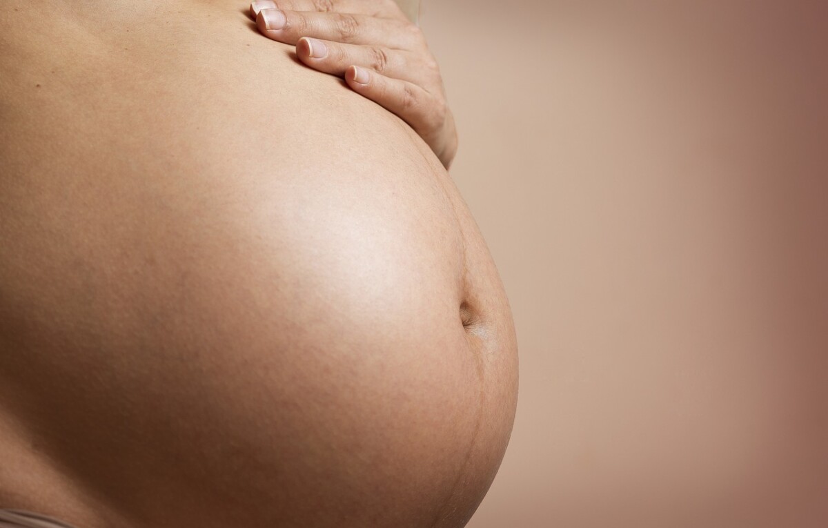
There are significant changes to hormone levels during pregnancy, particularly the hormones oestrogen, progesterone and relaxin. The increase in oestrogen during pregnancy enables the uterus and placenta to improve the formation of blood vessels, transfer nutrients from mother to baby and support the developing baby. Oestrogen levels steadily increase during pregnancy and reach their peak during the third trimester. Progesterone, in addition to relaxin, play a role in causing the laxity and loosening of ligaments and joints throughout the body. The release of these hormones enables the pelvis to widen to allow the baby to pass during childbirth. While the production and release of these hormones are critical for a pregnancy to continue and for childbirth, they can affect other structures, such as the pelvic floor.
Impact of Pregnancy and Birth on Pelvic Floor
The pelvic floor muscles are put under additional demand during pregnancy as they must support the weight of the growing baby, amniotic sac and placenta. The increasing downward pressure is one factor that can weaken the pelvic floor musculature. As mentioned above, altered hormonal levels also causes the muscles to become softened to allow the baby to birth, which can further weaken the pelvic floor. As a result, some women can experience an onset or increase in symptoms such urinary incontinence and urgency, pelvic organ prolapse and less commonly, faecal incontinence. For some women, incontinence and bladder/bowel weakness may continue after birth.
During labour and birth, there is further downward stress applied to the pelvic floor with active pushing. The pelvic floor muscles need to relax and the perineum needs to stretch to allow the baby to descend the vaginal canal. Depending on the size of the baby and the pelvic floor opening, the levator muscles must stretch 1.5 to more than 3 times their normal length. As a result, the pelvic floor muscles can be weakened or damaged in the process. The use of forceps or vacuum assistance during childbirth can further impact the pelvic floor.
It is commonly thought that having a c-section birth will prevent damage to the pelvic floor muscles. While there is less direct impact on the muscles, a c-section is a major abdominal surgery which requires incisions through multiple layers of abdominal muscle and tissue. Consequently, the core and pelvis in its entirety are affected. A c-section delivery also does not exclude the additional pressure endured by the pelvic floor throughout the nine months of pregnancy. Furthermore, c-section deliveries are often emergencies, which means that the woman has already had to actively push during labour, which contributes to the weakening of the pelvic floor.
Abdominal Separation
Diastasis rectus, also known as abdominal separation, occurs when the tissue known as the linea alba, which runs down the centre of the abdomen between the rectus abdominis muscles, becomes stretched and separates. This natural process commonly happens during pregnancy as the abdominal wall stretches to accommodate the growing foetus. The linea alba, being highly elastic, typically retracts after childbirth, allowing the muscles to come closer together again. However, in cases where the tissue is excessively stretched, the gap between the muscles may persist. Factors that increase the likelihood of developing diastasis rectus include multiple pregnancies, delivering larger babies, carrying twins or triplets, and being petite.
The prominence of diastasis rectus becomes more noticeable postnatally, once the abdominal wall is no longer under constant stretch. Typical signs include a visible bulge in the midsection, a soft or jelly-like feeling around the belly button, and a dome or cone shape around the belly button when the abdominal muscles are contracted.
Diastasis rectus itself is not typically painful, but women may experience weakness in their abdominals during daily activities such as lifting or sitting, as the muscles are not providing adequate support to the abdomen. This can lead to discomfort, particularly in the lower back, hips and pelvis.
Diastasis rectus can be diagnosed through palpation of the abdomen while the abdominal muscles are contracted. The health professionals will use their fingers to feel the abdominal area for gaps and changes in muscle tone. The separation is commonly recorded in finger widths, for example, two fingers of separation. In some instances, some health providers may use an ultrasound machine for a more accurate measurement. If you would like to check your abdominal separation at home, you can follow to steps below:
- Lie on your back with your knees bent
- Place your fingertips firmly above your belly button
- Lift your head and shoulders up off the floor to contract your abdominal muscles, while feeling for separation under your fingers. You should be able to feel the borders of the rectus muscles starting to squeeze your finger/s.
The primary approach to managing diastasis rectus involves activating and strengthening the abdominal muscles to encourage closure of the separation. However, it is crucial that exercises are prescribed and performed correctly. If exercises are too strenuous or performed incorrectly, they may exacerbate the separation and cause further issues such as visible doming or coning in the midsection. Therefore, targeted exercises should be tailored to the individual’s needs and capabilities, ensuring they support the gradual closure of diastasis rectus without causing unintended strain or complications.
It’s important to recognize that post-pregnancy, the abdominals may not fully return to their pre-pregnancy state of connection. This is a normal occurrence and should not cause undue concern for new mothers. The primary goal is for mothers to regain the ability to activate their abdominal muscles effectively and perform daily tasks without experiencing bulging or discomfort. The emphasis should be on functional strength and support rather than achieving a specific aesthetic outcome.
Return to Exercise
After pregnancy, the hormone relaxin can stay in your body for up to 4-6 months, leaving the ligaments and pelvic floor muscles more vulnerable to injury in the postnatal period. There will be reduced core stability and mechanical control as the abdominal muscles have naturally stretched during pregnancy and require time and care to recover, particularly following a caesarean section. For these reasons, it is important to increase exercise intensity slowly. There is also the risk of incontinence and/or pelvic organ prolapse if strenuous exercise is resumed too soon after birth when pelvic floor muscles haven’t fully recovered.
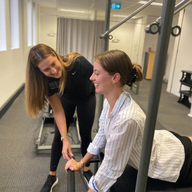
It is recommended that all women, regardless of how they deliver, seek out a pelvic health assessment with a specialist physiotherapist to evaluate strength, function and co-ordination of the abdominal and pelvic floor muscles which are often impacted by pregnancy and delivery. The importance of seeing a pelvic health physiotherapist is further highlighted if any of the following signs and symptoms are experienced:
- Heaviness / dragging in the pelvic area. These symptoms can be associated with prolapse.
- Leaking urine or inability to control bowel movements.
- Noticeable gap along the midline of your abdominal wall or doming of the abdomen with abdominal exercises.
- Pelvic or lower back pain.
- Ongoing or increased blood loss beyond eight weeks postnatal that is not linked to your monthly cycle.
Each woman’s return to an exercise program after childbirth varies and depends on several factors, including her prenatal exercise routine, the type of delivery and any associated trauma, the duration of the second stage of labor, the size of the baby, and the condition of her pelvic floor and abdominal muscles. However, it is generally recommended to follow a low-impact exercise regimen during the first three months postpartum, gradually incorporating higher-intensity exercises between three to six months postpartum, at the earliest.
In the initial days and weeks following childbirth, it is crucial to engage in exercises that gently activate the deep abdominal muscles and pelvic floor without excessively increasing intra-abdominal pressure. Gentle flat walking is also beneficial during this phase. The next phase involves gradually progressing abdominal challenges without doming/coning and incorporating general strengthening exercises for the upper and lower limbs. Functional exercises that mimic daily activities, such as lifting the baby or twisting to place the baby in a car seat, should also be introduced.
Higher-impact exercises like running or sports should only be resumed after thorough assessment and confirmation that abdominal and pelvic floor muscles have returned to their pre-pregnancy strength levels. As mentioned above, every return to exercise plan is individual, however a rough guide for return to run post baby is as follows:
Step 1 of your return to running plan is to get your pelvic floor run ready. If your pelvic floor doesn’t contract the way it should – every time your foot hits the ground, you will have a force of 1.6 to 2.5 times your body weight going through a pelvic floor which is unable to handle it. Before you run you should be able to activate your pelvic floor in standing as well as being able to do:
- 10x fast reps
- 8-12 reps of 6-8 second maximum voluntary contraction
- 60 seconds submaximal 30-50% contraction
The 2019 Returning to Running Postnatal Guidelines recommend that you should be able to achieve the following exercises without pain, heaviness, dragging or incontinence before starting running.
- Walking for 30 minutes
- Single leg balance 10 seconds
- Single leg squat 10 repetitions on each side
- Jog on the spot for 1 minute
- Forward bounds 10 repetitions
- Hop in place 10 repetitions on each leg
- Single leg ‘running man’: opposite arm and hip flexion/extension (bent knee) 10 repetitions on each side
Additionally, you should be able to complete 20 full repetitions of each of the following strength exercises:
- Single-leg calf raise
- Single-leg bridge
- Single-leg sit to stand
- Side-lying abduction
Weakness in these areas of strength testing should not be considered a barrier for return to running but instead identify where strength work can be directed.
In conclusion, it is clear that pregnancy exerts a significant impact on a woman’s body, both during gestation and childbirth, extending well into the postnatal phase. Understanding these effects and addressing common postnatal considerations is important. If you have any questions regarding this topic, please reach out to us at Bend + Mend. We are here to provide guidance and support during this time.
 References