 MRI (Magnetic Resonance Imaging)
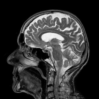
An MRI scan is a non-invasive scan that uses a powerful magnet and radio waves to create images of your tissues. During the scan you will be asked to lie on a moveable table that will slowly pass through the donut shaped scanner to image certain areas of the body.
The next part becomes more complex and harder to understand. Once within the “donut” hole a strong magnet is used to produce a magnetic field. This magnetic field forces the protons with in the body to align. A radiofrequency current is then pulsed though the body. Once this frequency is turned off the MR computer can analyse the energy released as the protons re-align and the time to realign. Generally, the faster these protons re-align different tissue the brighter the part shows in the image.
What is MRI used for?
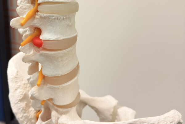
MRI provides good contrast images that are particularly good to view the non-bony or soft tissue parts of the body. The brain, spinal cord and nerves, as well as muscles, ligaments, and tendons can be seen more clearly on MRI than on CT or X-ray images.
What are the risks?
While the MR scan process sounds very complex, it does not use the potentially damaging ionizing radiation of x-rays or a CT. Because of this it is thought to be safe to use during pregnancy.
There are however some things that cannot go in the scanner. Because a large magnetic field is used, Pacemakers, implantable cardioverter-defibrillators, metal clips, metal valves or other metal implants can be dangerous because of the potential movement.
Claustrophobia can also be an issue within a MR scanner. The scan occurs in the enclosed space and it can take between 30 minutes to one hour. During this time, it is important to keep still so that the image does not blur.
Ultrasound scan
An ultrasound scan uses sound waves to produce two dimensional images of muscles, tendons, ligaments and joints. This is a non-invasive technique and it does not use ionising radiation.
To perform an ultrasound, scan a small probe is used alongside ultrasound gel. The probe transmits the high frequency sound waves through the gel and into the body. The sounds then bounce back though this probe and this information is collated by a computer to create an image.
What is it used for?
Musculoskeletal ultrasound is commonly used to look at tendons, ligaments and muscles.
What are the risks?
Because US scanning uses sound waves and does not use ionising radiation it does not have many risks. It does however have some limitations. Due to the depth a sound wave can penetrate, US is not give a clear image of deeper structures.
If you are not sure if you need a scan, or what type of scan you need, come and see us here at Bend + Mend in Sydney’s CBD. We will assess our injury and can advise you on the best course of management.
Click here to read Imaging Explain Part One: Xray and CT Scan