From professional athlete to weekend warrior, Anterior Cruciate Ligament (ACL) ruptures are a debilitating injury for all. Whilst ACL injuries have multiple mechanisms, low energy and non contact injuries are most common, accounting for 70% of ACL ruptures (Napolitano et al., 2024). An example of a non contact injury would be landing on uneven ground and the knee buckling inwards. When an ACL rupture occurs often patients will report an audible “pop” felt within the knee. Concurrent injuries to the medial meniscus, medial collateral ligament (MCL) are not uncommon as the mechanisms can be similar. The current research suggests that the incidence of non-contact ACL injuries can be reduced with interventions targeting ideal knee mechanics and replicating sport specific motions – that’s where physiotherapy comes in!
So why is this ligament so important?
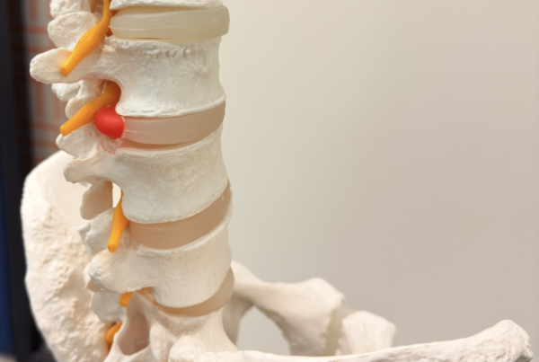
The ACL is important for knee stability. This ligament runs at an oblique angle from the back of the femur to the front of the tibia (thigh and shin bone respectively). The primary role of this ligament is to prevent anterior (forward) translation of the tibia or conversely posterior (backwards) translation of the femur. With movements such as lunging, jumping, landing, pivoting, and running seen with a variety of different sports – both contact and non contact.
What are Non -contact vs Contact ACL ruptures?
As mentioned above non-contact injuries make up 70% of ACL ruptures. The mechanism of injury involves dynamic knee valgus, tibial rotation and flexion of the knee. This is when the knee bends, drifts inwards creating a “knock knee” position and twists slightly inwards too. This is often seen in a jumping and landing motion or from a sudden deceleration and stop. Contact ruptures involve an external force – this is often from an opposing athlete. The knee collapses into forced valgus putting stress on the ACL because of this external force. This can be seen in tackles where players collide with one another.
Whilst the mechanism of non-contact ACL injury is common and consistent across disparate populations, certain groups show increased susceptibility due to a range of established and potential risk factors. Females are two to six times more likely to experience an ACL injury when compared to their male counter parts. This is due to the differences in anatomy. Typically, females have a smaller intercondylar notch (where the ACL attaches), a smaller ACL which has greater laxity subsequently being more susceptible to rupture (Parsons et al., 2021).
Improving biomechanics and correcting faulty moving patterns are useful in reducing risk of non contact ACL rupture. Working on jumping and landing mechanics and other sport specific movements should be implemented as part of an injury prevention programme. Your physiotherapist can help you develop a programme tailored to you and your sporting needs.
Risk by Sport
Certain sports have a great incidence of ACL ruptures amongst athletes. This is primarily due to the type of sport and the movement patterns that occur within these sports. A study of high school athletes outlined that the sports with the greatest risk for ACL rupture were soccer closely followed by basketball and volleyball (Joseph et al., 2013). When stratified by gender, girls’ soccer and basketball rated higher for injury risk when compared to their male counterparts. As suggested above, these injuries were comprised of more non-contact ruptures than contact ruptures. ACL ruptures are also very commonly seen in sports such as skiing and snowboarding. This is due to the quick change of direction required in these sports. It is also though that due to the stiffness of boots, combined with skis/snowboard facing in the opposite direction of your foot.
Within Australia specifically a rise in ACL injuries has been noted due to the fast paced and dynamic movements of AFL. Although the majority of athletes returned to play, the rate of rupture was significantly higher compared to other sports.
Conservative vs Surgical management
ACL ruptures can either be treated conservatively or through surgical management. Rodriguez et al. (2021) outlines that patient goals and levels of activity ultimately direct whether conservative or surgical treatment is necessary. Conservative management involves a strict structured rehabilitation programme to support the ACL deficient knee. With surgical management a similar rehabilitation programme is adopted to improve outcomes and healing of the new ACL and facilitating a successful return to play.
The surgical management of the ACL typically involves reconstruction using grafts, with hamstring tendons and the patellar tendon being the two most common options. Each graft type has distinct advantages, disadvantages, and indications based on the patient’s specific needs.
Hamstring Tendon Graft
The hamstring tendon graft is harvested from the semitendinosus (medial hamstring) and gracilis tendons. This technique is favoured for several reasons. First, it involves less postoperative pain compared to patellar tendon harvesting, as the patellar tendon approach can be more invasive and result in anterior knee pain. Secondly, the hamstring graft offers excellent biomechanical properties, including strength and elasticity, which are vital for knee stability. The surgery is performed arthroscopically (keyhole), with the graft being threaded through bone tunnels created in the femur and tibia, secured with fixation devices such as screws or buttons.
However, there are some downsides. Hamstring tendon harvesting may result in muscle weakness and a slight reduction in hamstring strength, which can affect athletic performance. Additionally, there is a potential for graft elongation over time, which can impact stability if not monitored carefully.
Patellar Tendon Graft
The patellar tendon graft involves taking a central portion of the tendon along with bone blocks from the patella and tibia. This graft type is known for its robust fixation and a low rate of graft failure, making it a popular choice among athletes, especially those involved in high-demand sports. The rigid nature of the bone-tendon-bone construct provides excellent stability, and the patellar tendon has a high tensile strength, ensuring effective load distribution across the knee.
However, this approach is not without its challenges. Harvesting the patellar tendon can lead to anterior knee pain, especially during kneeling or squatting activities, and may have longer recovery times compared to hamstring grafts. There is also a risk of patellar fracture or patellar tendinopathy post-surgery.
Regardless of graft type, a well-structured rehabilitation program is essential for optimal recovery. Early stages focus on controlling swelling and regaining range of motion, progressing to strength training and sport-specific drills. Athletes typically return to competitive activities within 6 to 12 months, with outcomes generally being positive if those are adherent.
Conservative management is thought to have a poorer prognosis when considering return to sport and participation due to the crucial role of the ACL in cutting, pivoting and contact sports. However, decisions on management need to be individualised to the patient and their desired goals and their current impairment with their ACL deficient knee.
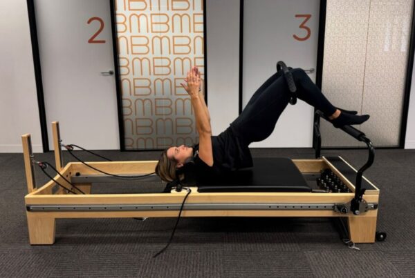
Physiotherapy and rehabilitation following ACL rupture
Whether a surgical or conservative approach has been decided. Both rehabilitation plans adopt similar features. Improving neuromuscular control, strength around the knee and surrounding muscle groups such as the quadriceps and hamstring muscles, adopting good biomechanics and focusing on sport specific movements that are meaningful to the client and their participation goals.
A well-structured postoperative rehabilitation program is crucial for successful recovery following anterior cruciate ligament (ACL) reconstruction. The program aims to restore knee function, strength, and stability while minimizing the risk of complications such as stiffness or reinjury. Typically, the rehabilitation process is divided into several phases, each with specific goals and activities.
Phase 1: Immediate Postoperative Care (0-2 Weeks)
The first phase focuses on pain management, reducing swelling, activating the quadriceps and regaining range of motion. Patients are encouraged to elevate the leg and apply ice to manage swelling. Weight-bearing is usually allowed, depending on the surgeon’s protocol. Sometimes if a subsequent meniscal tear has been repaired or debrided weight bearing status may be altered. Use of crutches are case by case but are often encouraged in these early stages to encourage correct gait patterns.
Physiotherapy begins with gentle passive and active range-of-motion exercises to prevent stiffness especially around knee extension (straightening the knee). Quadriceps activation exercises are also introduced to improve neuromuscular control and activation.
Exercises may include:
- Isometric knee extension – where the back of the knee is pushed into the ground activating the quadriceps.
- Heel slides – sitting on a chair and sliding the heel under the chair to promote knee flexion.
As mentioned above reducing swelling in this early phase is important to reduce arthrogenic muscle inhibition (AMO). This is when there is swelling present within and around a joint and the chemicals within this swelling inhibit muscle activation – thus reducing the ability for strong contraction.
Phase 2: Early Rehabilitation (2-6 Weeks)
In this phase, the focus shifts to increasing range of motion and beginning light strengthening exercises. Patients typically work on achieving at least 90 degrees of knee flexion in this period. Gradual weight-bearing is encouraged, often transitioning to full weight-bearing as tolerated. Closed kinetic chain exercises are encouraged in this phase. These are exercises where the foot or body part is fixed to a stationary point.
Exercises may include:
- Wall slides / mini squats
- Glute/hamstring bridges
- Calf raises
Closed kinetic chain exercises help strengthen the quadriceps without placing excessive stress on the knee.
Returning to day-to-day activities and resuming “normal” tasks such as walking and driving are introduced in this period through the guidance of your physiotherapist.
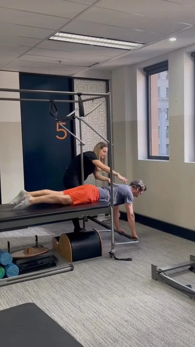
Phase 3: Strengthening and Functional Training (6-12 Weeks)
During this phase, the rehabilitation program intensifies, focusing on strength, proprioception, and functional movements. Resistance training, including exercises targeting the quadriceps, hamstrings, and hip muscles, are integrated. Balance exercises are reintroduced as they are crucial for improving proprioception. Patients are typically encouraged to return to low-impact activities like cycling or swimming. Single leg exercises are also introduced at this phase also.
Exercises may include:
- Leg press and leg extension machines
- Balance exercises on mats or completing dual tasks
- Single leg squats
Outcome measures are often used within this phase to track progress and appropriateness to progress to the next phase of strengthening. I.e. having a poor single leg squat it wouldn’t be safe to progress to a hop or a single leg plyometric exercise.
Phase 4: Advanced Strengthening and Return to Sport (3-9 Months)
At this stage, the focus is on building strength, agility, and sport-specific skills. Patients engage in higher-intensity exercises, plyometrics, and agility drills, such as ladder drills and cone sprints. The goal is to restore hopping abilities (technique, distance and endurance), return to agility and sports specific modified drills, and to regain full strength and balance.
Exercises may include:
- Ladder drills
- Box jumps
- Change of direction, cutting and pivoting drills
Phase 5: Return to Full Activity (9+ Months)
By nine months post-surgery, some patients are ready to return to sports provided they meet specific criteria, including strength symmetry and functional tests. A gradual return to competitive activities is encouraged, with ongoing emphasis on conditioning and injury prevention strategies.
Psychological readiness is another huge factor when considering returning to sport. There are many questionnaires designed to assess this including the Anterior Cruciate Ligament-Return to Sport after Injury (ACL-RSI).
Rehabilitation is crucial for restoring strength, stability, and functionality to the knee after an ACL rupture. Proper rehabilitation not only facilitates the return to sport safely but also minimises the risk of re-injury and long-term joint problems. Your physiotherapist will help you through this tailored structured programme giving you the confidence to get back to the activities most meaningful to you.
References