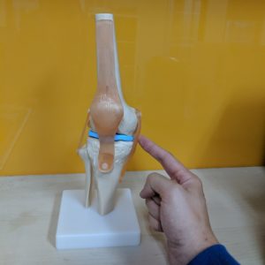
 The football season is well underway, and the list of injuries continues to grow. Knee injuries are often some of the more gruesome sporting injuries to observe, and often can lead to an extended period of time on the sideline, if not season ending.
Statistically speaking, ligament injuries are the most commonly reported type of injury of the knee. The ACL (Anterior Cruciate Ligament) and MCL (Medial Collateral Ligament) are the most commonly injured ligaments of the knee. The MCL is injured in at least 42% of all knee ligament injuries, and isolated MCL injuries make up 29% of all knee ligamentous injuries.
The Medial Collateral Ligament is one of the four main ligaments of the knee. It is a broad, flat band located on the inner (medial) aspect of the knee. It originates on the medial femur (inner aspect of the thigh bone), crosses the knee joint and attaches onto the medial tibia (inner aspect of the shin bone). The primary function of the MCL is to provide stability to the medial aspect of the knee, preventing valgus movement of the knee (the knee collapsing inwards).
MCL Anatomy:
The MCL is composed of a superficial and a deep layer. The superficial layer runs from just posterior to the medial femoral epicondyle down 6 cm beyond the medial tibial plateau. The deep layer is a thickening of the joint capsule.
The MCL resists valgus forces that would push the knee medially (inwards).
Injuries to the MCL:
If the knee falls into a rapid valgus position (rolls or is pushed in), often due to contact (tackle, impact) or non-contact (poor change of direction, poor landing/stepping biomechanics), this will stress the MCL, causing damage to the ligament.
MCL injuries are often sustained in combination with other structures of the knee, which adds to the complexity of the management. In this blog we will be discussing the management of isolated MCL injuries, with no other structures affected.
Rehabilitation following an MCL injury:
Luckily the MCL has very high healing potential, therefore a majority of MCL injuries are managed non-operatively. The MCL’s high healing potential is due to a good blood supply, which facilitates a strong healing response. However, premature resumption of sports/activity may stress the healing ligament too soon and lead to chronic instability and pain.
Ligament injuries are graded on a scale of 1-3, from least to most severe. The time-frames of these injuries can be seen below.
Grade 1: Minor Tear – 0-2 weeks
Pain along ligament, no instability or gapping of joint with stress testing.
Grade 2: Partial Tear, approximately 50% of the fibres – 2-6 weeks
Partial gapping (5-10 mm) of joint with stress testing at 30 degrees of knee flexion.
Grade 3: Complete Rupture – 6-12+ weeks
Wide gapping (>10 mm) of joint at 30 degrees of knee flexion (no end feel).
Management of the more severe injuries are treated in a hinged knee brace set to allow 30-90 degrees of motion. This prevents full knee extension, holding the knee in a stable position without stressing the MCL while it is healing. It is imperative that throughout the time in the brace the knee does not extend fully until the ligament has healed, in severe injuries this is approximately 6 weeks.
Rehabilitation following immobilisation will focus on restoring range of motion, strength, landing mechanics, agility, plyometrics and power. Usually in that order.
At Bend + Mend in Sydney’s CBD our Physio’s can provide you with an elite rehabilitation program for a speedy and fast return to pre-injury activities so feel free to book in today.
