Stress fractures are a common overuse injury, especially among active individuals involved in high impact sports like running, jumping, and athletics. In this post, we’ll explore what stress fractures are, who is most at risk, how they develop and what one might expect to see when managing these sorts of injuries.
Types of Stress Fractures
Firstly, there are two types of stress fractures. The main being fatigue fractures. These occur in otherwise healthy bone which has been subject to abnormal or excessive repetitive stress. Common in athletes or those that are engaged with endurance-based sports; this type arises from overload not underlying bone weakness. In contrast insufficiency fractures occur when a normal stress is applied to a weakened bone. This can be due to a variety of different reasons but most commonly with individuals with osteoporosis of other metabolic diseases. These conditions can be read about in one of our previous blog posts.
Epidemiology
As mentioned, stress fractures are a common sports injury and account for 10-20% of all sports injuries according to the current literature. Of these a staggering 95% occur in the lower limb. This again is due to the weight bearing nature of joints and bones. Of these lower limb injuries stress injuries to the tibia (shin bone) account for 50%, the most common being medial tibial stress syndrome known as shin splints. Bones in the foot known as tarsals account for 25%. The bones in the forefoot known as the metatarsals, the thigh bone known as the femur and the outside of the shin known as the fibula account for roughly 9% each. Females are 2-4 times more likely to suffer stress fractures than their male counterparts. This is often due to an overall lower bone mineral density, hormonal fluctuations (especially post-menopausal) and general biomechanical differences such as less bone mass and alignment. Adolescents and younger adults aged 15-25 are often most at risk of fatigue fractures and this increases with involvement in high-impact sports.
Aetiology
So why might stress fractures happen? The aetiology of a pathology refers to the risk factors involved. Intrinsic factors are things refer to internal factors, these often (but not always) cannot be drastically changed. One’s bone density, muscular strength, hormonal status, genetics are all examples of intrinsic factors. Extrinsic factors are things external that can influence risk. How often one is training, the type of footwear they wear, rest and recovery are all examples of extrinsic factors. Understanding these factors is crucial for early diagnosis, prevention and management of bone stress injuries.
Pathophysiology – What Happens Inside the Bone
In simple terms stress fractures occur when repetitive cyclic loading creates micro-damage in the bone which is outpaced by the body’s ability to repair it. The bone is repeatedly loaded i.e. from running, this micro-damage accumulates in the cancellous bone (porous bone under the outer layer). Activity increases in the bone cells called osteoclasts. These are responsible for bone resorption and uptake of older bone cells. Osteoblast cells which form new bone cannot keep up with the resorption, thus an overall weakening of the bone occurs. This can lead to a stress reaction, a stress fracture or at worst a full fracture if left untreated.
Wolff’s law was developed by German anatomist Julius Wolff that describes bone remodeling regarding stress and impact on bone. With mechanical stress bone remodels and adapts to this stress thus getting strong. However, without rest the process becomes maladaptive.
Imaging
Magnetic Resonance Imaging (MRI) is the gold standard for imaging stress reactions and fractures. This is due to the ability to detect mild changes in the bone before they become full fractures. Early changes to both the cortical (hard outer) and the cancellous bone (porous inner) can be identified on MRI. X-ray unfortunately cannot always pick up on stress reactions. There are a multitude of different ways to classify stress fractures. Ultimately higher stages indicate worse prognosis.
The Fredericson classification utilises MRI imaging to grade stress injuries.
- Grade 1: Periosteal oedema only (inflammation in the outermost layer of the bone)
- Grade 2: Bone marrow oedema (T2 only) (inflammation in soft tissue within cancellous bone)
- Grade 3: Bone marrow oedema on T1 and T2
- Grade 4: Visible fracture line
T1-weighted images show fat as bright and water as dark, making them ideal for visualising anatomy, while T2-weighted images show both fat and water bright, with water appearing brighter, which is excellent for detecting inflammation and fluid.
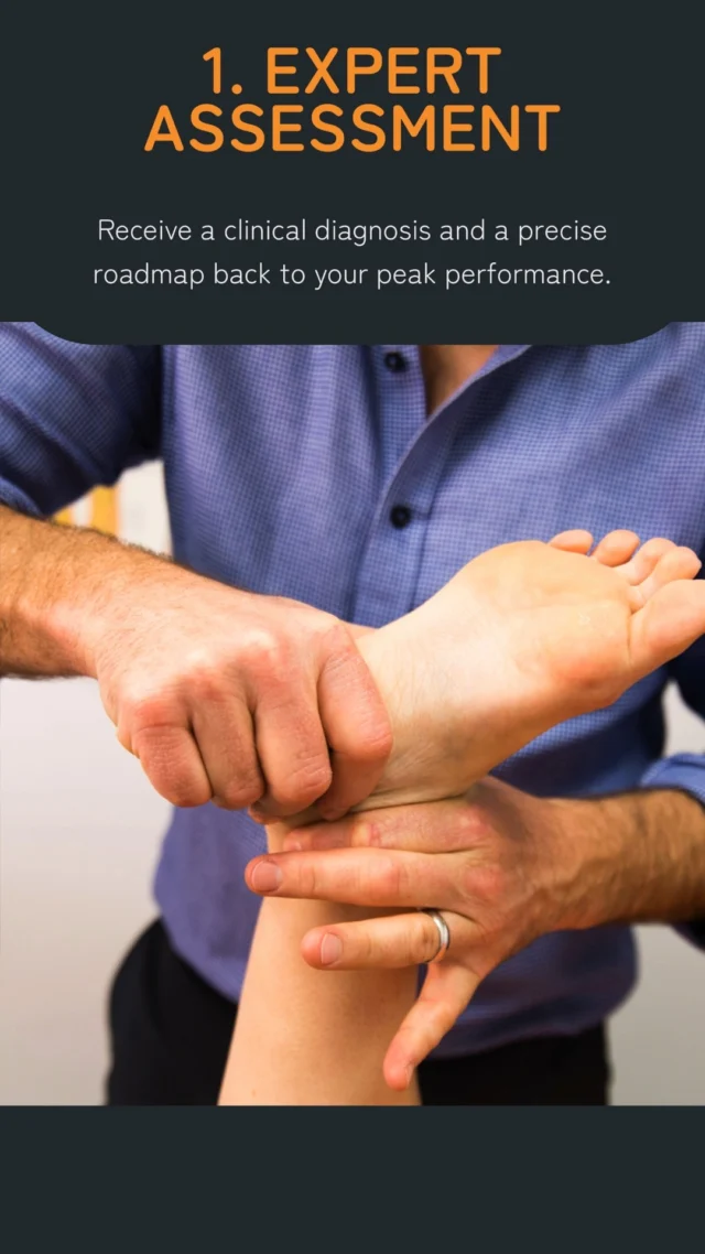
Clinical Tests:
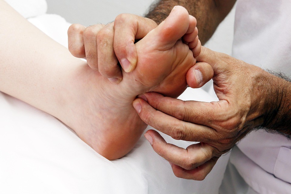 Physiotherapists use a variety of different tests and tools to help identify stress reactions and fractures to help guide management and treatment.
In the current literature the diagnostic tests used to identify stress injuries are not considered strong and are varied across the board. Focal palpation remains one of the stronger testing methods meaning if a client has a positive result (pain on palpation of the injured site) we can rule in confidently the possibility that a stress injury may be the diagnosis. Other tests include using tools such as a tuning fork to vibrate the injured area in which a positive result would be pain reproduction; this however only yields moderate diagnostic value according the literature.
Ultimately a comprehensive subjective history (including analysis of risk factors) and clinical reasoning remain the strongest tool. So don’t be alarmed if your physio is asking you a lot of questions!
Management:
Managing these injuries can be broken up into three phases: acute, sub-acute and return to play. Each phase has different goals and objectives.
Acute phase:
This is when you may present to the physiotherapist with symptoms such as pain, inability to weight bear and complete the activities meaningful to you such as running, walking and jumping. Within this phase the goals are to reduce pain, identify the bony stress and to promote healing. Your physiotherapist will give you advice on how to modify your activity to reduce load to the injured bone. Depending on severity and location of your injury a moonboot may be needed. Again, your physiotherapist can help with this process.
Sub-acute phase:
Once pain starts to decrease and pain levels are under control you can commence the next phase of treatment. Within this phase gradual reloading of the bone can take place. This might involve weaning out of a moonboot if one was prescribed or starting to get back to some lower impact activities such as walking. Biomechanical and strength deficits can be addressed within this phase also to prevent re-injury / future injury.
Return to play phase:
Lastly return to play. The goal is for successful safe reintegration into full activity. This can be guided by pain-free symptoms or even with clear diagnostic imaging. Structured programmes to gradually introduce load can be used such as walk/run programmes and all modifiable risk factors should be addressed to ensure safe return to sport and play.
Conclusion:
Early identification to allow for adequate load management is crucial in the treatment of bone stress related injuries. This identification comes from both identification of risk factors as well as a few key diagnostic tools. Rehabilitation should be phased, progressive, and individualised; your Physiotherapist will help guide you through this process.
Early intervention and structured rehab can make a world of difference in outcomes and get you back to doing the tasks you love!